Spinal decompression is defined.
Spinal decompression refers to a group of surgical procedures used to alleviate painful compressed or pinched spinal nerves (neural impingement). When the spinal canal and nerve apertures become too tiny owing to bulging or collapsed discs, swollen joints, loosened ligaments, or bony growths, they might become inflamed. Compression on the spinal nerve roots causes leg pain.
Spinal decompression surgery is commonly used to treat the following conditions:
- Spinal stenosis is a narrowing of the spinal column that puts pressure on the nerves around it.
- Sciatica occurs when a diseased spinal disc impinges on a nerve below the disc.
- Spinal injuries can result in fractures and tissue edoema.
- Metastatic spinal cord compression can result from cancer spreading from one part of the body, such the lungs, by pressing on the spinal cord or nerves.
Overview of the Spine
33 small bones, known as vertebrae, that are stacked one on top of the other and separated by a supple intervertebral disc make up the spine. The annulus fibrosus, which is made up of several collagenous layers and resembles an onion, surrounds the nucleus pulposus, a soft jelly-like substance located in the centre of the intervertebral disc. This disc joins the vertebrae, facilitates movement between them, and acts as a shock absorber for the spine. The spinal cord and nerve roots are therefore shielded by the vertebrae.
Who Needs a Spinal Decompression Procedure?
Spinal decompression surgery is indicated for patients with spinal stenosis. Spinal stenosis, a condition in which the spinal canal narrows, can cause ongoing pain, numbness, and muscle weakness in the arms or legs. The degenerative changes that cause the ligaments to thicken and the facet joints to widen are what cause this condition, which typically affects the elderly.
Those who require spinal decompression surgery are suggested to have the following conditions:
- Anxiety, numbness, weakness, or pins and needles in the legs or feet
- Leg pain that is worse in the back
- Inability to respond to any type of medication or physical therapy results in problems standing or walking, which might affect one's quality of life.
Spinal decompression procedure
To treat the symptoms of nerve decompression, a variety of surgical techniques are often performed, including:
- Microdiscectomy Sciatica, a lumbar herniated disc that causes leg pain, is frequently treated with micro decompression. The procedure comprises using microsurgical techniques to cut off the piece of the disc pressing against the nerve.
- Lumbar Laminectomy: A lumbar laminectomy with open decompression is frequently used to address leg pain brought on by lumbar spinal stenosis. After the removal of the bony roof lamina and any thickened ligaments that were covering it, the left and right sides of the spinal canal will be totally decompressed.
- Laminotomy: This surgery involves preserving the majority of the lamina while removing a small portion of the ligaments and laminar roof that are located above the spinal canal. Either the left or right side of the spinal canal can be decompressed using it. This method preserves the lamina's natural support, which lowers the risk of postoperative spinal instability. An endoscope may occasionally be used, allowing for a less intrusive incision.
Each of these actions can be taken singly or in combination, depending on the urgency and requirements of the circumstance.
Before the procedure
- Several diagnostic procedures are performed before to the treatment, including electrocardiograms, chest X-rays, and blood tests.
- The patient's medical history is documented, including any allergies, prescriptions taken, vitamins taken, previous procedures, bleeding patterns, and anaesthetic reactions.
- The patient should stop using all non-steroidal anti-inflammatory medicines (NSAIDS) and blood thinners one week before surgery (such as Coumadin and aspirin).
- Also, the patient is advised to stop smoking, chewing tobacco, and drinking alcohol one week prior to the surgery and for two weeks following it because these behaviors may induce bleeding problems.
The patient is admitted to the hospital on the day of the procedure. Moreover, avoiding eating or drinking after midnight the night before surgery is advised. An intravenous line is put into the arm.
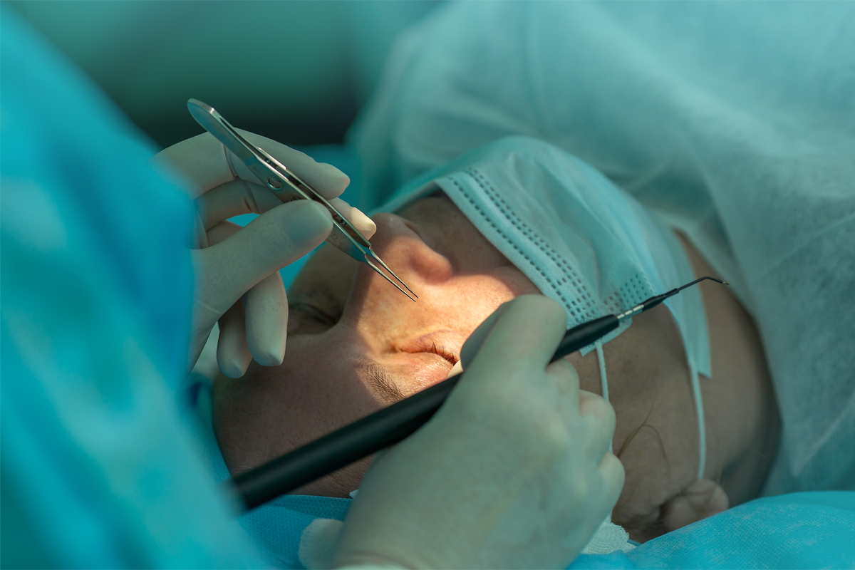
All during the procedure
General anaesthesia is utilised during surgery, and the entire procedure might take anywhere between one and three hours, depending on the complexity.
- For the operation, the patient is prepared.
- Around the centre of the back, a cut is made across the pertinent vertebra.
- The back muscles are split down the middle and pushed to either side to show the lamina of each vertebra.
- After obtaining an X-ray of the exposed bone to determine which vertebrae need to be operated on, the appropriate procedure—a laminectomy or laminotomy—is performed.
- Decompress the spinal nerve by performing a procedure known as a foraminotomy, which involves trimming or cutting the facet joints, which are situated right above the nerve roots, to create more space for the latter.
After the procedure
The patient will stay in the hospital for four or five days, depending on the procedure. To relieve their postoperative agony, the patient will keep taking painkillers. Following the surgery, the patient can sip on liquids before gradually moving on to a full meal. Four to five days after the procedure, the patient can take a shower.
Throughout the first several weeks, the patient might require assistance with basic everyday chores like getting dressed and taking a shower. Fatigue after the procedure is common. One can gradually get back to their normal routine.
After being hospitalized
The patient should observe the following limitations after the procedure:
- It is advised that the patient refrain from operating a motor vehicle for two to four weeks after surgery or until the doctor provides the all-clear.
- Should not sit for prolonged periods of time.
- Should not lift anything heavy or bend or twist at the waist.
- The patient is allowed to do any housework and yard chores following the initial follow-up consultation. Mopping, vacuuming, ironing, loading and unloading the dishwasher, washing and drying clothes, and gardening all included in this.
- The client is advised to give up smoking. Smoking hinders recovery by increasing the risk of complications including infections and inhibiting bone fusion.
- The patient is instructed to walk.


Comments
Post a Comment